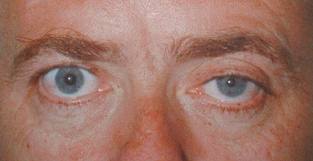
Imagine a world where your eye and facial muscles are disrupted, leaving you with a mysterious condition that affects your appearance and vision. Welcome to the intriguing realm of Horner’s Syndrome, a rare but fascinating condition that has puzzled medical professionals for years. From the drooping of the eyelid to the constricted pupil and lack of sweating on one side of the face, Horner’s Syndrome presents a unique set of symptoms. But what exactly causes this syndrome? How is it diagnosed and treated? And what does it mean for your overall prognosis? Prepare to unravel the mysteries of Horner’s Syndrome as we explore its anatomy, etiology, evaluation, and much more.
Definition and Overview
What is the definition and overview of Horner Syndrome? Horner Syndrome, also known as Bernard-Horner syndrome, is a rare condition characterized by partial ptosis (drooping of the upper eyelid), miosis (constriction of the pupil), and facial anhidrosis (lack of sweating on one side of the face). It can be acquired or congenital, and it is named after Johann Friedrich Horner, who described it in 1869. The syndrome is caused by the interruption of sympathetic fibers along their pathway, which can occur at various locations, such as intracranially, intraorbitally, or extracranially. The epidemiology of Horner Syndrome shows a frequency of 1 per 6,250 population, and it can occur at any age and in any ethnicity. The pathophysiology involves abnormalities in the sympathetic innervation of the eye, resulting in symptoms such as ptosis, miosis, and anhidrosis. Differential diagnosis includes other conditions that present similar symptoms, such as Adie pupil, physiologic anisocoria, and iris sphincter muscle damage. Diagnostic criteria include a detailed medical history, physical examination, and various tests, such as pupillary measurements, imaging studies, and pharmacological testing. Understanding the causes, epidemiology, pathophysiology, differential diagnosis, and diagnostic criteria of Horner Syndrome is crucial for accurate diagnosis and management of this rare condition.
Anatomy and Physiology
The sympathetic innervation of the eye plays a crucial role in the development of Horner Syndrome. The anatomy and physiology of Horner Syndrome involve the disruption of the sympathetic pathway that innervates the eye. Here are five key points about the anatomy and physiology of Horner Syndrome:
- Sympathetic innervation involves three neurons: First-order neurons originate from the hypothalamus and terminate at the spinal cord. Second-order preganglionic neurons synapse in the superior cervical ganglion. Third-order postganglionic fibers innervate sweat glands and blood vessels of the face.
- Interruption of sympathetic fibers can occur extracranially, intracranially, or intra-orbitally, leading to Horner Syndrome.
- The causes of Horner Syndrome can be categorized based on the location of the disruption, with preganglionic Horner Syndrome often associated with pulmonary malignancies.
- Symptoms of Horner Syndrome include partial ptosis (drooping of the upper eyelid), miosis (constriction of the pupil), anhidrosis (lack of sweating), and flushing and sweating abnormalities on the affected side of the face.
- Diagnostic tests and evaluation for Horner Syndrome may include pupillary diameter measurement, examination of the eyelids for ptosis, imaging studies, and pharmacological testing like the topical cocaine test or apraclonidine test.
Understanding the anatomy and physiology of Horner Syndrome is essential for accurate diagnosis and appropriate management of the condition.
Etiology and Risk Factors
To understand the etiology and risk factors of Horner Syndrome, it is important to consider the underlying causes that can disrupt the sympathetic pathway involved in the condition. Horner Syndrome can occur due to various causes and predisposing factors. Common etiologies include tumors, such as lung cancer, as well as swollen lymph glands in the neck, dissection of the aorta, and thoracic aortic aneurysm. These conditions can interrupt the sympathetic fibers that connect the eyes and brain, leading to the development of Horner Syndrome. Other risk factors include trauma or surgical intervention that affects the second-order preganglionic neurons in the sympathetic pathway. In some cases, Horner Syndrome may be congenital, meaning it is present at birth. The underlying conditions that contribute to Horner Syndrome can vary, and the severity and symptomology of the syndrome depend on the location and degree of denervation. Understanding the pathogenesis and triggers of Horner Syndrome is crucial for accurate diagnosis and management of the condition.
Clinical Presentation and Symptoms
Patients with Horner Syndrome typically present with a combination of clinical features and symptoms that include partial ptosis, miosis, and facial anhidrosis. These manifestations result from disruption of sympathetic innervation to the eye. The following clinical presentation and symptoms may be observed:
- Partial ptosis: Drooping of the upper eyelid, which may be mild to moderate.
- Miosis: Constriction of the pupil, leading to a smaller pupil size on the affected side.
- Facial anhidrosis: Decreased sweating on the affected side of the face.
- Apparent enophthalmos: The eye may appear sunken due to the ptosis and miosis.
- Increased amplitude of accommodation: The ability to focus on near objects may be enhanced.
It is important to note that the severity and symptomology of Horner Syndrome can vary depending on the location and degree of sympathetic denervation. Prompt diagnosis and appropriate treatment are crucial for optimal management. The underlying cause of Horner Syndrome should be identified and addressed. While the long-term prognosis of idiopathic Horner Syndrome is generally benign, complications may arise from the underlying pathology. Regular follow-up and monitoring of symptoms are essential to ensure proper care and patient education should focus on recognizing the possible presentations of Horner Syndrome and understanding the importance of exploring the underlying cause.
Diagnostic Tests and Evaluation
After discussing the clinical presentation and symptoms of Horner Syndrome, the next step is to evaluate and diagnose the condition through diagnostic tests. The evaluation begins with a detailed history and medication review to rule out any potential miotic or mydriatic agent use. Localization of the lesion is crucial for determining the underlying cause and guiding management. Balance, hearing, and swallowing problems may indicate a more central process, while prior trauma or surgical intervention suggest involvement of second-order neurons. Imaging modalities such as chest X-ray, CT scan, MRI, and ultrasonography can help identify the location and extent of the lesion. Pharmacological testing, including the topical cocaine test, hydroxyamphetamine test, and apraclonidine test, can also be used to assess sympathetic innervation. In congenital cases, urine tests may be performed to rule out neuroblastoma. These diagnostic tests and evaluations play a crucial role in identifying the underlying causes of Horner Syndrome and guiding appropriate management strategies.
Treatment Options and Management
Treatment for Horner syndrome depends on the underlying cause and may involve a combination of medical, surgical, and supportive interventions. The following options are available for the management of Horner syndrome:
- Medical treatment: This may include medications to address specific underlying conditions, such as tumors or infections. Medications can also be used to manage symptoms like ptosis or miosis.
- Surgical intervention: In some cases, surgical procedures may be necessary to correct the underlying cause of Horner syndrome. This may involve removing tumors or repairing damaged nerves.
- Specialist consultations: Depending on the underlying cause, consultations with specialists such as neurologists, ophthalmologists, or otolaryngologists may be required to provide comprehensive care.
- Follow up and monitoring: Regular follow-up appointments are important to assess the progress of treatment and monitor any changes in symptoms. This allows for adjustments in the treatment plan if needed.
- Addressing the underlying cause: Identifying and addressing the underlying cause of Horner syndrome is crucial for effective treatment. This may involve further diagnostic tests or consultations with other specialists.
It is important to work closely with healthcare professionals to determine the most appropriate treatment options based on your specific condition and needs.
Prognosis and Complications
The prognosis of Horner syndrome is generally favorable, with most patients experiencing either improvement or resolution of their symptoms. The long-term outlook for patients with Horner syndrome depends on the underlying cause of the condition. Complications are related to the specific etiology of Horner syndrome. For instance, if the syndrome is caused by a tumor, the potential risks include tumor growth and spread. In cases where the syndrome is congenital, there may be no significant complications or risks. However, regular follow-up care is essential to monitor for any changes in symptoms or potential complications. Patient outcomes vary depending on the individual and the specific cause of Horner syndrome. Some patients may see a complete resolution of their symptoms, while others may experience ongoing mild symptoms. It is important for patients to work closely with their healthcare providers to determine the appropriate management and follow-up care to optimize their prognosis.
Patient Education and Support
Patient education and support play a crucial role in managing Horner syndrome and understanding its underlying causes and management options. It is important for patients to have access to accurate information and resources to cope with the challenges of living with this condition. Here are some key factors to consider when it comes to patient education and support:
- Patient education: Providing patients with information about Horner syndrome, including its symptoms, causes, and treatment options, can empower them to make informed decisions about their care. This may involve explaining the anatomy and physiology of the condition, as well as discussing the different diagnostic tests and imaging studies that may be used to evaluate Horner syndrome.
- Support groups: Connecting patients with support groups or online communities can offer a sense of belonging and provide a platform for sharing experiences, tips, and coping strategies. Support groups can also be a valuable resource for emotional support and practical advice on managing the daily challenges associated with Horner syndrome.
- Coping strategies: Teaching patients coping strategies, such as relaxation techniques and stress management skills, can help them navigate the emotional and psychological impact of living with Horner syndrome. Encouraging open communication and fostering a supportive environment can also contribute to better coping outcomes.
- Treatment options: Educating patients about the different treatment options available for Horner syndrome, such as medication, surgical interventions, and lifestyle modifications, can help them make informed decisions about their care. It is important to explain the potential benefits and risks associated with each treatment option, as well as any potential side effects or complications.
- Lifestyle modifications: Providing guidance on lifestyle modifications, such as protecting the affected eye from sunlight and wearing sunglasses, can help patients manage their symptoms and prevent further complications. It is important to emphasize the importance of regular follow-up appointments and monitoring to ensure optimal management of Horner syndrome.


