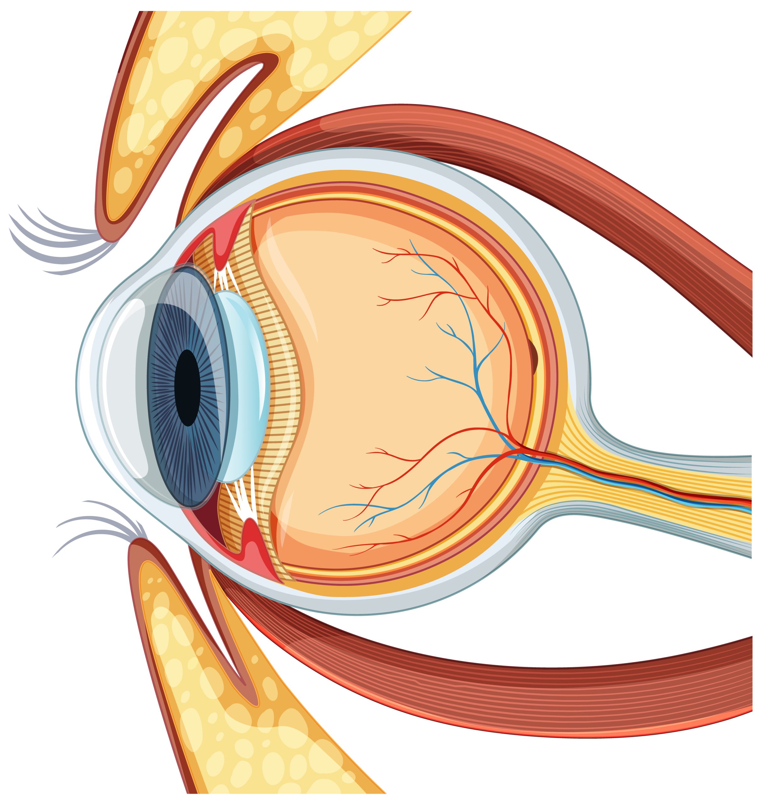
Are you ready to explore the intricate world of external ocular muscles? Look no further! In this comprehensive guide, we’ll take you on a journey through the depths of these enigmatic muscles and unveil their crucial role in eye movement and visual perception. Whether you’re a medical professional looking to enhance your understanding or a student embarking on an anatomy journey, this article is designed to provide you with a wealth of knowledge. So join us as we unravel the mysteries of external ocular muscles and gain a deeper understanding of their significance in the oculomotor system.
Importance of External Ocular Muscles
Understanding the importance of external ocular muscles can greatly enhance your knowledge of eye movement disorders and guide the development of effective treatments. The external ocular muscles play a crucial role in controlling eye movements and are essential for visual perception. These muscles are commonly targeted in surgical interventions for strabismus and other ocular motility disorders. Therefore, a comprehensive understanding of their structure and function is vital for predicting their response to interventions and developing new treatments.
Clinical assessment of the external ocular muscles is essential for evaluating their functionality and detecting any abnormalities. By testing different eye movements, such as those controlled by the medial rectus and lateral rectus muscles, clinicians can assess the function of specific muscles and diagnose eye movement disorders.
The importance of external ocular muscles is further highlighted in the context of eye disorders. For example, in myasthenia gravis, these muscles are affected by afferent signals from various brain regions, such as the cerebellum and vestibular nuclear complex. Structural analysis of extraocular muscles has provided valuable insights into the pathology of myasthenia gravis, including the examination of myosin heavy chain isoform patterns and the study of ultrastructural relationships between muscle growth and peripheral nerve autografts.
Overview of External Ocular Muscles
Take a closer look at the external ocular muscles and their functions. The comparative anatomy of external ocular muscles reveals that there are six muscles responsible for controlling eye movement. These muscles include the superior rectus, inferior rectus, lateral rectus, medial rectus, superior oblique, and inferior oblique muscles. Each muscle has specific functions, such as elevation, depression, adduction, abduction, intorsion, extorsion, and elevation of the eyelid.
Age-related changes in external ocular muscles can occur, leading to alterations in muscle function and eye movement. These changes may include a decrease in muscle strength and flexibility, which can affect binocular vision. The external ocular muscles play a crucial role in binocular vision by coordinating the movement of both eyes, allowing for depth perception and accurate visual perception.
Exercise has been shown to have positive effects on external ocular muscles. Regular eye exercises can help improve muscle strength, flexibility, and coordination, leading to better eye movement control and visual acuity. It is important to note that specific exercises targeting the external ocular muscles may be beneficial for individuals with certain eye conditions or muscle imbalances.
Additionally, the external ocular muscles also play a role in facial expressions. These muscles work in conjunction with other facial muscles to produce various expressions, such as raising the eyebrows, squinting, or widening the eyes. The coordinated action of the external ocular muscles contributes to the overall facial expression and nonverbal communication.
Functions of External Ocular Muscles
The functions of the external ocular muscles encompass the precise control and coordination of eye movements, enabling visual perception and depth perception. These muscles play a crucial role in maintaining eye alignment, allowing both eyes to work together and focus on objects of interest. They also contribute to the ability to track moving objects and shift gaze from one point to another. The functions of the external ocular muscles are affected by various factors, including extraocular muscle disorders and the effects of aging on these muscles. To study the external ocular muscles, imaging techniques such as MRI and ultrasound can be used to visualize their structure and function. In cases of external ocular muscle injuries, therapeutic approaches may include physical therapy, surgery, or the use of medication to manage inflammation and promote healing. Understanding the functions of the external ocular muscles is essential for diagnosing and treating eye movement disorders and maintaining optimal visual function.
Innervation and Blood Supply of External Ocular Muscles
To understand the innervation and blood supply of external ocular muscles, it is important to examine the cranial nerve innervations and vascular sources that provide essential support to these muscles. The cranial nerve innervations responsible for controlling the extraocular muscles are the oculomotor nerve (CN III), trochlear nerve (CN IV), and abducens nerve (CN VI). The oculomotor nerve innervates the superior rectus, inferior rectus, medial rectus, inferior oblique, and levator palpebrae superioris muscles. The trochlear nerve controls the superior oblique muscle, while the abducens nerve controls the lateral rectus muscle.
In terms of blood supply, the extraocular muscles receive their vascular support from the branches of the ophthalmic artery. The ciliary arteries, which are branches of the ophthalmic artery, provide blood to the recti muscles. Each rectus muscle, except for the lateral rectus, receives blood from two anterior ciliary arteries. The lateral rectus muscle is supplied by only one ciliary artery. The inferior rectus and inferior oblique muscles are supplied by branches of the infraorbital artery.
In a clinical eye exam, the functionality and movement of the extraocular muscles can be assessed. Different eye movements, such as those involving the medial rectus and lateral rectus muscles, can be tested to evaluate the function of specific muscles. Mnemonics can also be used to remember muscle actions and movements. Overall, understanding the innervation and blood supply of the external ocular muscles is crucial for the diagnosis and treatment of various eye disorders.
Clinical Examination of External Ocular Muscles
When examining the external ocular muscles, it is important to assess their functionality and movement in order to diagnose and treat various eye disorders. To conduct a clinical examination of external ocular muscles, several techniques and diagnostic tests can be employed. These include:
- Visual assessment: The examiner visually inspects the eyes for any abnormalities in alignment, size, and movement. This can help identify common eye muscle disorders such as strabismus or nystagmus.
- Ocular motility assessment: The examiner assesses the range of eye movements in different directions using the “H test” or other eye movement tests. This helps evaluate the function of specific muscles, such as the medial rectus and lateral rectus muscles.
- Imaging advancements: Advancements in eye muscle imaging, such as MRI or ultrasound, can provide detailed information about the structure and function of the external ocular muscles. This can aid in the diagnosis and treatment planning for various eye muscle disorders.
Treatment options for eye muscle disorders depend on the specific condition and may include corrective lenses, eye exercises, or surgical interventions. By utilizing clinical examination techniques, diagnostic tests, and advancements in eye muscle imaging, healthcare professionals can accurately diagnose and effectively treat common eye muscle disorders.
Learning Anatomy of External Ocular Muscles With Kenhub
Learn about the anatomy of external ocular muscles with Kenhub, a comprehensive online resource that provides high-quality illustrations, articles, and interactive learning tools. Kenhub offers a range of advantages for students studying the anatomy of external ocular muscles. With curated learning paths, you can easily navigate through the material and focus on the specific topics you need to learn. The high-quality illustrations and articles provide detailed information about the structure and function of each muscle, allowing you to develop a thorough understanding of their anatomy.
To further enhance your learning experience, Kenhub offers a free 60-minute trial of Kenhub Premium, which gives you access to additional resources such as videos, quizzes, and galleries on various anatomy topics. This interactive approach allows you to actively engage with the material and reinforce your knowledge.
In addition to learning the basic anatomy, Kenhub also covers topics related to extraocular muscle disorders, ocular motility interventions, and extraocular muscle adaptations. This comprehensive coverage ensures that you have a well-rounded understanding of the subject.
Kenhub is a valuable tool for anyone studying the anatomy of external ocular muscles. Its user-friendly interface, high-quality illustrations, and interactive learning tools make it an excellent resource for anatomy learning. Whether you are a medical student, healthcare professional, or anatomy enthusiast, Kenhub provides the resources you need to master the anatomy of external ocular muscles and excel in your studies.
Role of External Ocular Muscles in Eye Disorders
Understanding the role of external ocular muscles in eye disorders is essential for diagnosing and treating various conditions. These muscles play a crucial role in eye movement and visual perception. Here are some key aspects to consider:
- Ocular proprioception and efference copy: These mechanisms are involved in registering visual direction and have implications for eye movement disorders. They contribute to the coordination and control of eye movements.
- Thyroglobulin independent cytotoxicity: Studies have observed this phenomenon in human eye muscle cells, particularly in the context of Graves’ ophthalmology. It indicates the involvement of immune-mediated processes in eye disorders.
- Visual localization after strabismus surgery: The treatment of strabismus, a condition characterized by misalignment of the eyes, can impact visual localization. Understanding the underlying mechanisms can improve surgical outcomes and patient satisfaction.
- Expression of thyrotropin receptor mRNA: Research has focused on studying the expression of this mRNA in healthy and Graves’ disease retro-orbital tissue. It provides insights into the pathogenesis of Graves’ ophthalmology.
Myasthenia Gravis and External Ocular Muscles
If you have myasthenia gravis, you may experience weakness and fatigue in your external ocular muscles. Myasthenia gravis is an autoimmune disorder that affects the neuromuscular junction, leading to muscle weakness and fatigue. The effects of medication on myasthenia gravis can vary, with some medications helping to improve muscle strength and others having minimal effect. The role of extraocular muscles in strabismus, or misalignment of the eyes, is crucial. These muscles control the movement and coordination of the eyes, and any weakness or dysfunction can result in strabismus. Ultrastructural changes in the extraocular muscles have been observed in individuals with myasthenia gravis, including alterations in the neuromuscular junction and muscle fiber degeneration. Genetic factors also play a role in the development of myasthenia gravis, with certain genes increasing the risk of developing the condition. Therapeutic approaches for myasthenia gravis include the use of immunosuppressive medications, such as corticosteroids and immunomodulatory drugs, to reduce inflammation and improve muscle function. Other treatments include plasmapheresis and intravenous immunoglobulin therapy, which help to remove antibodies and provide temporary relief from symptoms. Overall, understanding the effects of medication, the role of extraocular muscles, ultrastructural changes, genetic factors, and therapeutic approaches is essential for managing myasthenia gravis and maintaining optimal ocular function.
Structural and Functional Analysis of External Ocular Muscles
Examine the structural and functional analysis of external ocular muscles to gain insights into their properties and mechanisms of action.
- Effects of aging on external ocular muscles: As we age, the external ocular muscles may weaken, leading to difficulties in eye movements and vision stability. These changes can contribute to age-related vision problems.
- Role of external ocular muscles in vision stability: The external ocular muscles play a crucial role in maintaining the stability of our vision. They work together to ensure that the eyes are aligned and can focus on objects accurately.
- Impact of exercise on external ocular muscle strength: Regular exercise, including eye exercises, can help strengthen the external ocular muscles. This can improve their functionality and contribute to better eye movement control and overall visual health.
- Relationship between external ocular muscles and dry eye syndrome: Dysfunction in the external ocular muscles can contribute to dry eye syndrome. The muscles may not be able to properly distribute tears across the surface of the eye, leading to dryness and discomfort.
- Role of external ocular muscles in refractive errors: Refractive errors, such as nearsightedness or farsightedness, can occur due to imbalances in the external ocular muscles. These imbalances can affect the shape of the eye and its ability to focus light accurately on the retina.
Understanding the structural and functional analysis of external ocular muscles is essential for diagnosing and treating various eye conditions and maintaining optimal visual health.
Neural Connections of External Ocular Muscles
How do the neural connections of external ocular muscles contribute to their coordination and precise control of eye movements? The neural connections of external ocular muscles play a crucial role in coordinating and controlling eye movements. These connections are responsible for transmitting signals from the brain to the muscles, allowing for the precise activation and relaxation of specific muscles to achieve desired eye movements.
The neural pathways of eye movement involve the cranial nerves, specifically the oculomotor nerve (CN III), trochlear nerve (CN IV), and abducens nerve (CN VI), which innervate the different external ocular muscles. These nerves ensure the proper activation and coordination of the muscles involved in eye movements.
Neurological disorders affecting eye muscles can disrupt these neural connections, leading to impaired eye movement control. Conditions such as myasthenia gravis can affect the transmission of signals between nerves and muscles, resulting in weakness and fatigue of the eye muscles.
Furthermore, extraocular muscle development and plasticity are influenced by neural connections. During development, neural signals guide the growth and organization of these muscles, ensuring their proper function. Additionally, the role of proprioception, the sense of body position and movement, is essential in eye muscle function. Proprioceptive feedback from the extraocular muscles helps in maintaining eye alignment and coordinating eye movements, contributing to their precise control. Overall, the neural connections of external ocular muscles are integral to their coordination, plasticity, and functioning in both normal and pathological conditions.
Surgical Interventions for External Ocular Muscles
To perform surgical interventions for external ocular muscles, you will need to understand the specific techniques and procedures involved. Here are some key points to consider:
- Surgical outcomes: The success of the surgery is typically determined by the alignment of the eyes and the improvement in ocular motility. Surgical interventions aim to correct strabismus and other ocular motility disorders, leading to improved visual function and cosmesis.
- Complications: While rare, complications can occur during or after surgery. These may include infection, bleeding, scarring, or damage to surrounding structures. Careful surgical planning and meticulous technique can help minimize these risks.
- Advancements: Over the years, there have been significant advancements in surgical techniques and instrumentation. These include the use of adjustable sutures, minimally invasive approaches, and improved imaging technologies. These advancements have contributed to better surgical outcomes and patient satisfaction.
- Postoperative care: After surgery, patients require close monitoring and follow-up visits to ensure proper healing and alignment. Eye drops or ointments may be prescribed to prevent infection and reduce inflammation. Physical therapy or vision exercises may also be recommended to optimize functional outcomes.
- Patient satisfaction: The ultimate goal of surgical interventions is to improve the patient’s quality of life and overall satisfaction. Assessing patient satisfaction involves evaluating visual function, cosmetic appearance, and the patient’s subjective experience postoperatively.
Future Directions in Understanding External Ocular Muscles
Exploring new research avenues is essential for gaining a deeper understanding of external ocular muscles and their complexities. Future research in this field should focus on elucidating the underlying mechanisms of muscle function and the development of therapeutic approaches for various pathological conditions affecting the external ocular muscles. Experimental techniques such as advanced imaging modalities, molecular biology, and genetic studies can provide valuable insights into the structure and function of these muscles.
One area of future research could involve investigating the molecular signaling pathways involved in the development and maintenance of external ocular muscles. By understanding these pathways, researchers can identify potential targets for therapeutic interventions. Additionally, studying the role of stem cells in muscle regeneration and repair can open up new avenues for treating muscle-related disorders.
Another important direction for future research is to explore the pathophysiology of ocular motility disorders and their impact on the external ocular muscles. Investigating the changes in muscle structure and function in conditions such as strabismus, myasthenia gravis, and dysthyroid eye disease can provide valuable insights into the underlying mechanisms and help develop targeted treatment strategies.
Furthermore, the development and refinement of experimental techniques, such as in vivo imaging and biomechanical analysis, can contribute to a better understanding of the dynamics and biomechanics of external ocular muscles. These techniques can help unravel the intricate interplay between muscle contraction, eye movement, and visual perception.


